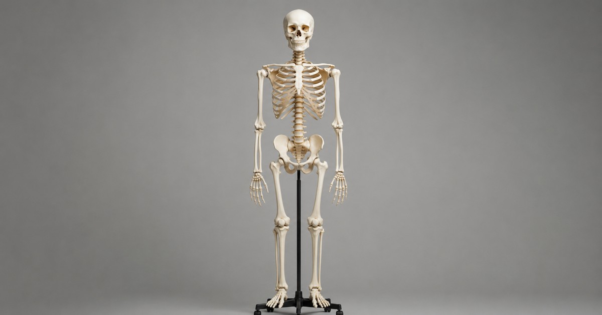
How many bones are in the human body? The number most people learn in school is 206. That is the standard count for a healthy adult. But the full answer is more interesting than a single number.
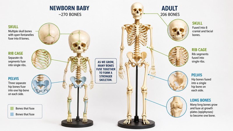
Babies are born with roughly 270 to 300 bones. Some adults have as few as 206 and others have as many as 213. The count changes throughout your entire life, peaks in infancy, then drops steadily through childhood and adolescence before stabilizing in your mid-twenties. After that, the number stays the same but the bones themselves never stop changing.
How many bones are in the human body? Adults typically have 206 bones. Newborns have between 270 and 300. The difference exists because many of the separate bones you are born with gradually fuse into single larger bones as you grow. By your mid-twenties, fusion is complete and you carry the same 206 bones for the rest of your life.
Your skeleton does far more than hold you upright. It produces 2 million red blood cells every second inside your bone marrow, stores most of the calcium your body needs to function, protects every major organ, and continuously rebuilds itself in response to the forces you place on it. The bones inside you right now are not the same bones you had ten years ago. The skeleton replaces approximately 10 percent of its tissue every year, giving you a new skeleton roughly every decade.
How Many Bones Are in the Human Body: The Direct Answer
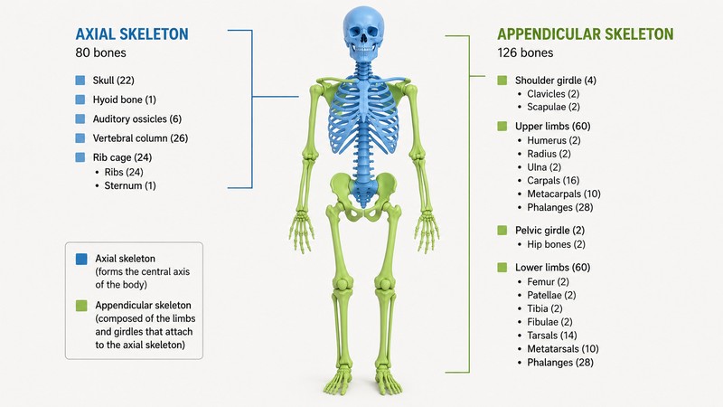
The standard adult count is 206 bones. Johns Hopkins Medicine breaks this down precisely: 80 bones form the axial skeleton, which includes the skull, spine, sternum, and ribs, and 126 bones form the appendicular skeleton, which includes the arms, shoulders, wrists, hands, legs, hips, ankles, and feet.
That count of 206 does not include teeth, which are not classified as bones, and does not include sesamoid bones, which are small bones embedded within tendons rather than connected to the main skeleton. The most well-known sesamoid bone is the patella, the kneecap, which is actually included in the 206 count. Other smaller sesamoid bones in the hands and feet are not.
The actual range for healthy adults is 206 to 213, because some people naturally have extra bones. Some individuals are born with an extra pair of ribs, known as cervical ribs. Some have additional small bones in their spine or extra bones in their wrists and feet called accessory bones. None of these variations cause health problems, and many people never know they have them.
Why Babies Have Nearly 100 More Bones Than Adults
The reason newborns have so many more bones than adults has everything to do with birth and early development.
A baby must pass through the birth canal, which requires the skull to compress and flex during delivery. This is only possible because the infant skull is not a single rigid structure. It is five separate bone plates separated by fibrous gaps called fontanelles, the soft spots you can feel on a newborn’s head. These plates can overlap slightly during delivery, reducing the skull’s diameter enough to pass through. After birth, the plates gradually fuse together. The fontanelles close within the first one to two years of life.
Beyond the skull, a newborn’s entire skeleton contains far more cartilage and far fewer mineralized bone segments than an adult. The pelvis, which in adults is a single basin-shaped structure, begins as three separate bones in infants that do not fully fuse until the late teens. The coccyx, the tailbone, begins as four or five separate segments that fuse into one. The sacrum, at the base of the spine, is five vertebrae at birth that become one bone by adulthood.
Cleveland Clinic pediatrician Dr. Matthew Badgett explains that having more, smaller, softer bones gives babies the flexibility needed both for delivery and for rapid early growth. Flexible bones bend rather than break, protecting infants during the accident-prone first years of life. As the skeleton solidifies and the child grows larger and heavier, fused, harder bones provide the structural strength needed to support increasing body weight.
How Bone Fusion Works: Ossification Explained
The process that converts the cartilage and separate bone plates of a newborn into the 206-bone adult skeleton is called ossification. It begins before birth and is not complete until your mid-twenties.
Ossification happens in two ways. In endochondral ossification, cartilage models are gradually replaced by bone tissue. Specialized cells called osteoblasts deposit calcium and phosphate minerals into the cartilage framework, converting it into hard bone. This is how the long bones of the arms and legs form and grow during childhood. The growth plates at the ends of these bones remain cartilage until growth is complete, then ossify and fuse, ending the possibility of further height gain.
In intramembranous ossification, bone forms directly from connective tissue without a cartilage intermediate. The flat bones of the skull form this way, which is why newborns have those membranous fontanelles rather than cartilaginous ones.
UC Davis bone biology research identifies three types of specialized cells that carry out ossification and ongoing bone maintenance throughout life: osteoblasts, which build new bone tissue; osteoclasts, which break down and remove old bone tissue; and osteocytes, which are mature bone cells embedded in the bone matrix that regulate the remodeling process and detect mechanical stress. These three cell types work in coordinated cycles throughout your entire life, not just during development.
The Two Divisions of the Human Skeleton
Anatomists divide the 206 adult bones into two groups based on their position and function.
The axial skeleton: 80 bones
The axial skeleton runs along the central axis of the body. It consists of the skull, the vertebral column, the sternum, and the ribs.
- Skull: 22 bones in total, including 8 cranial bones that protect the brain and 14 facial bones. In adults all are fused except the mandible, the lower jaw, which remains movable.
- Vertebral column: 26 bones including 7 cervical vertebrae in the neck, 12 thoracic vertebrae in the mid-back, 5 lumbar vertebrae in the lower back, the sacrum, and the coccyx. The sacrum and coccyx are each formed by the fusion of multiple vertebrae during development.
- Thoracic cage: 25 bones including the sternum and 24 ribs arranged in 12 pairs. The first seven rib pairs connect directly to the sternum. The next three connect via cartilage shared with the rib above. The last two are floating ribs with no anterior attachment.
The appendicular skeleton: 126 bones
The appendicular skeleton consists of the limbs and the structures that attach them to the axial skeleton. The word appendicular comes from the Latin for ‘to hang from.’
- Upper limbs: 64 bones total, including the shoulder girdles, the humerus of the upper arm, the radius and ulna of the forearm, and 27 bones in each hand counting the carpals, metacarpals, and phalanges.
- Lower limbs: 62 bones total, including the pelvic girdle, the femur of the thigh, the patella, the tibia and fibula of the lower leg, and 26 bones in each foot.
The hands and feet together account for more than half of all the bones in the body. Each hand contains 27 bones and each foot contains 26. The reason is the complexity of fine motor control and locomotion. More individual bones allow more precise articulation across more joints.
The Smallest and Largest Bones in the Body
The range in bone size within a single human body is extraordinary.
The femur, the thigh bone, is the longest, heaviest, and strongest bone in the body. It runs from the hip joint to the knee and supports the entire weight of the upper body during standing, walking, and running. UC Davis reports that the femur can withstand forces up to 30 times a person’s body weight. In a 70-kilogram adult, that is a compressive strength of approximately 2,100 kilograms before fracturing under ideal conditions.
The stapes, one of the three tiny bones in the middle ear, is the smallest bone in the body. It measures approximately 3 millimeters in length, roughly the size of a grain of rice. The stapes works alongside the malleus and incus to transmit sound vibrations from the eardrum to the fluid of the inner ear. All three ossicles together, the ossicular chain, amplify sound by a factor of approximately 22 before it reaches the cochlea.
Between the femur and the stapes, the 206 bones of the adult skeleton range from structures that can withstand the force of a car crash to structures smaller than the head of a pin, all performing precise mechanical functions.
What Your Bones Actually Do: 6 Functions Most People Do Not Know
1. Structural support and movement
Bones provide the rigid framework that holds the body upright and gives muscles something to pull against. Without bones, muscle contraction would produce no directed movement. Tendons attach muscles to bones at precise insertion points that act as levers, translating muscle force into controlled movement of joints.
2. Protection of vital organs
The skull encloses and protects the brain. The vertebral column protects the spinal cord. The rib cage surrounds and shields the heart and lungs. The pelvis protects the bladder, reproductive organs, and lower digestive structures. Each major organ in the body has a dedicated bony enclosure built around it.
3. Blood cell production
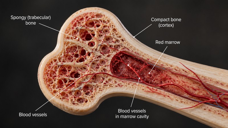
Red bone marrow, found primarily in the flat bones including the sternum, ribs, vertebrae, and pelvis, produces all blood cells through a process called hematopoiesis. The marrow generates approximately 2 million red blood cells every single second throughout your life. It also produces white blood cells for immune defense and platelets for clotting. Without healthy bone marrow, the immune system and oxygen delivery systems both fail rapidly.
4. Mineral storage and release
Bones store 99% of the body’s calcium and 85% of its phosphate. These minerals are not permanently locked in place. The skeleton releases calcium into the bloodstream whenever blood calcium levels drop below the required threshold, and absorbs calcium from the blood when levels are too high. This makes the skeleton the primary regulator of blood calcium, which controls muscle contraction, nerve function, and heart rhythm. Osteoporosis, which weakens bones as they age, is partly a consequence of the skeleton releasing more calcium than it replaces over time.
5. Fat storage
Yellow bone marrow, which replaces red marrow in the long bones of the limbs during late childhood and adolescence, consists primarily of fat cells. This fat serves as an energy reserve. During prolonged starvation or extreme metabolic stress, the body can draw on yellow marrow lipids for fuel. Yellow marrow can also partially convert back to red marrow if the body urgently needs more blood cell production.
6. Endocrine function
Research has established that bones are endocrine organs, not just structural ones. Osteocalcin, a hormone produced by osteoblasts during bone formation, circulates through the bloodstream and influences insulin secretion, muscle performance during exercise, brain development, and male fertility. This was largely unknown until the early 2000s and remains an active area of research. The skeleton is not a passive support structure. It actively communicates with the rest of the body through hormonal signaling.
How Your Bones Rebuild Themselves Throughout Your Life
Your skeleton is not a fixed structure. It is in a constant state of controlled demolition and rebuilding called bone remodeling.
Osteoclasts continuously dissolve and remove sections of old or micro-damaged bone. Osteoblasts follow behind them, depositing new bone tissue to fill the cleared space. The process operates across the entire skeleton simultaneously, with different regions on different remodeling cycles. Approximately 10 percent of the skeleton is replaced each year in healthy adults, meaning the skeleton you have today is substantially different from the one you had a decade ago.
Remodeling serves several functions. It repairs microscopic damage from daily mechanical stress before cracks can grow into fractures. It adjusts bone density and shape in response to changing load patterns. Athletes in weight-bearing sports develop denser, thicker bones in the limbs they use most. Astronauts in zero gravity, with no mechanical load on their skeletons, lose bone density rapidly without countermeasures because the remodeling cycle tips toward removal without the stimulus of load to drive formation.
Medical News Today’s review of bone biology notes that remodeling also regulates calcium and phosphate concentrations in the blood. When blood calcium drops, parathyroid hormone signals osteoclasts to dissolve more bone and release calcium into the circulation. When calcium is adequate, calcitonin slows osteoclast activity. The skeleton functions as a calcium bank, constantly making withdrawals and deposits to keep blood chemistry in balance. This process connects to the broader chemistry of the human body, where multiple organ systems regulate the same minerals through different mechanisms.
Why the Number of Bones Can Vary Between Adults
The standard answer of 206 assumes a typical adult skeleton. A meaningful fraction of people have a slightly different count, and most never know it.
Cervical ribs are extra ribs that grow from the seventh cervical vertebra in the neck rather than from the thoracic vertebrae where ribs normally attach. They occur in approximately 0.5 to 1 percent of people and are usually harmless, though in some cases they compress nerves or blood vessels and cause symptoms.
Accessory ossicles are extra small bones that sometimes form in the wrists, ankles, or feet when areas of cartilage that would normally fuse completely instead ossify as separate bones. They are relatively common, found in roughly 10 to 20 percent of people depending on the specific location, and are almost always asymptomatic.
Sutural bones, also called Wormian bones, are extra bones that occasionally form within the sutures of the skull. They appear as small additional bone islands within the fibrous joints between the major skull bones and are considered anatomical variants rather than abnormalities.
On the other end, some people have fewer than 206 bones if certain fusions that are normally incomplete have occurred more extensively, or if they were born without expected bones in their fingers or toes through a condition called oligodactyly.
How Bone Count and Bone Strength Change With Age
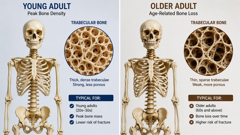
After bone fusion is complete in the mid-twenties, the number of bones stays at 206 for the rest of life. The quality and density of those bones, however, follows a predictable arc.
Bone density peaks between ages 25 and 35. During this window, the remodeling cycle is balanced, with formation and resorption occurring at roughly equal rates. After 35, resorption gradually begins to outpace formation in most people. Bone density decreases slowly but steadily.
In women, estrogen plays a protective role in bone density by suppressing osteoclast activity. The hormonal changes of menopause cause a significant acceleration in bone loss during the first five to ten years after menopause, with some women losing up to 20 percent of their bone density in this period. This is the primary reason women have substantially higher rates of osteoporosis than men.
In men, testosterone provides a similar protective effect. The more gradual decline in testosterone with age produces a slower, more linear bone density loss compared to the post-menopausal acceleration in women.
Physical activity, particularly weight-bearing exercise such as walking, running, and resistance training, is the strongest behavioral stimulus for maintaining bone density through adulthood. Mechanical load signals osteocytes, which communicate with osteoblasts to increase bone formation in loaded areas. Sedentary behavior removes this stimulus and allows resorption to dominate.
Frequently Asked Questions
How many bones are in the human body at birth?
Newborns have between 270 and 300 bones, with the exact number varying by gestational age at birth and individual development. The range exists because some of the cartilaginous structures counted as separate bones at birth are so early in their development that different researchers classify them differently. By any count, newborns have roughly 100 more bones than adults.
At what age do you have 206 bones?
Most people reach the adult count of 206 bones by their mid-twenties, though the process can continue until age 25 or occasionally slightly later. The final fusions to complete are usually the growth plates of the long bones and the clavicle, which is often the last bone to finish ossifying.
Which part of the body has the most bones?
The hands and feet combined account for more than half of all the bones in the body. Each hand has 27 bones and each foot has 26, giving the four extremities a total of 106 bones, more than half the full adult count. This reflects the mechanical complexity required for fine motor tasks in the hands and for the balance and propulsion demands placed on the feet.
Can you break all 206 bones?
Technically yes, though some bones are far more resistant than others. The femur, the strongest bone in the body, typically requires massive force to fracture, the kind produced by high-speed vehicle accidents or severe falls. The small bones of the hands, wrists, and feet fracture far more easily. Stress fractures in foot bones are common in runners and military recruits. Some bones, like the bones of the inner ear, rarely fracture because they are enclosed in dense protective tissue and never experience direct impact.
Do bones grow back after being broken?
Yes. Bone is one of the few tissues in the body capable of complete regenerative healing rather than scar repair. After a fracture, new blood vessels grow into the break site within days. Specialized cells produce a soft callus of cartilage that bridges the gap. Osteoblasts then replace the cartilage callus with new bone tissue through the same ossification process that built the skeleton originally. Complete healing can take weeks to months depending on which bone broke and how severe the fracture was.
How do bones relate to the immune system?
Red bone marrow produces all the cells of the immune system, including lymphocytes, neutrophils, and macrophages, through hematopoiesis. The skeleton is therefore the primary manufacturing site for immune defenses. Conditions that damage bone marrow, such as certain cancers and autoimmune diseases, directly compromise immune function. Bone marrow transplants are used as treatments for severe immune deficiencies and certain blood cancers because they introduce healthy stem cells capable of rebuilding both the blood cell and immune cell supply. This close relationship between bone and immunity connects to the extraordinary facts about how the human immune system operates, including defenses that most people never think about.
The One-Paragraph Answer
The adult human body has 206 bones, divided into 80 in the axial skeleton and 126 in the appendicular skeleton. Newborns have between 270 and 300, with the extra bones fusing gradually through childhood, adolescence, and into the mid-twenties as ossification converts cartilage to bone and separate plates merge into single structures. Some adults naturally have 207 to 213 bones due to extra ribs, accessory ossicles, or sutural skull bones. The skeleton is not a passive framework. It produces 2 million red blood cells every second, stores 99% of the body’s calcium, protects every major organ, secretes hormones that influence insulin and muscle function, and rebuilds approximately 10% of itself every year through the coordinated action of bone-building and bone-dissolving cells. The number of bones in your body has not changed since your mid-twenties. The bones themselves have been completely rebuilt multiple times since then.